
Magnetic Resonance Imaging (MRI)
Table of Contents
What is MRI?
“Magnetic Resonance Imaging (MRI) a method of using magnetic field to produced detailed image of every internal structure of human body.”
For many years, magnetic techniques aided chemical analysis in food and petrochemical industries. The development of large – bore homogeneous magnets and computer assisted imaging ( as a CT scanning ) .
How MRI works ?
When a substance is placed in magnetic field, spinning protons within the nuclei act like small magnets and align themselves within field . A superimposed electromagnetic pulse (radio wave) at a specific frequency displaces the hydrogen protons. The transverse component of the magnetization vector generates the MRI signals .These signals are used to display as image .
The T1 component( or spin lattice relaxation) depends on the time taken for the protons to realign themselves with the magnetic field and reflects the proton return to thermal equilibrium which interact with surrounding lattice.
The T2 component ( Spin- Spin relaxation) is time taken for the protons to return to their original ‘out of phase ‘ state and depends on the locally ‘energized’ protons.
Interpretation of abnormal MRI image
Any structural abnormalities and abnormal intensities which show a change in tissue T1 or T2 weighting in term to normal grey and white matter . ( A prolonged T1 relaxation time in MRI gives hypo intensity, that is more black and a prolonged T2 relaxation time in MRI gives hyperintensity that is more white) .
Usefulness of MRI in cardiac condition
- In cardiac cycle the MRI scans need to be ‘gated’ which allow the scanner to produce moving
images of the heart and mediastinal structures - MRI is very helpful in imaging the aorta.
- It is also useful for detecting and evaluation of the right ventricle that
is difficult to image by echocardiography. - MRI help in selecting patients for revascularization procedures, or in
identifying those with the myocardial infiltration, such as that seen
with the sarcoid heart disease and arrhythmogenic right ventricular
cardiomyopathy.
Usefulness of MRI in kidney and urinary tract
Magnetic resonance imaging (MRI) offers excellent resolution
and gives good distinction . It’s very useful for local staging of prostate, bladder and penile cancers. Magnetic resonance angiography (MRA) provides an alternative to the CT for imaging renal vessels but involves administration of gadolinium-based contrast media, which may carry risks for patients with impaired renal function. MRA gives good images of main renal vessels.
Usefulness of MRI in bone diseases
Magnetic resonance imaging (MRI) gives detailed information on anatomy, allow 3D visual image of bone and soft tissues that cannot be adequately seen in plain X-rays. The technique is valuable in the diagnosis and treatment of many musculoskeletal diseases.
T1-weighted sequences are useful for defining anatomy
T2-weighted sequences are the useful for assessing tissue water content.
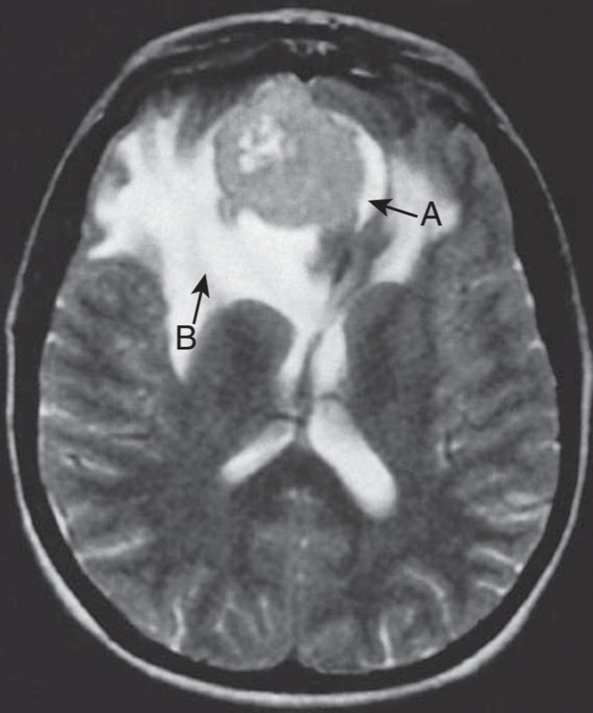
Usefulness of MRI in neurological condition
MR Angiography is a procedure used to evaluate blood flow in various artery and veins in brain and spinal cord . Mainly used for aneurysms in brain and vascular malformation . Some of the MRI used for neurological condition are
- Diffusion – weighted MRI (DWI): Image based on transitional movement of water and small molecules within the brain.
- Perfusion -weighted MRI(PWI):Image obtained by ‘bolus tracking’ after rapid contrast injection
- Functional MRI (fMRI)
- Magnetic resonance spectroscopy (MRS)
Advantage of MRI
- MRI can directly scan any plane e.g. coronal, sagittal , oblique
- No ionizing radiation is used in MRI .
- MRI is highly sensitive to any change in tissue e.g. demyelination plaques .
- No bone artifacts, e.g. intracanalicular acoustic neuroma.
Disadvantage of MRI
- In MRI , Limited Slice thickness -2-3 mm with 3 Tesla; 3-5 mm with 1.5 Tesla (CT-1mm)
- Bone imaging is limited to display of marrow.
- The patient may feel Claustrophobia.
- MRI Cannot use with pacemaker or ferromagnetic implant patient .
- It is Expensive then any other imaging technique .
- Less widely available
- MRA images blood flow in various vessels of body .
Recent Post