
Tumour
Table of Contents
Definition of Tumors
A mass of tissue formed as a result of abnormal, excessive, uncoordinated, autonomous and purposeless proliferation of the cells even after cessation of stimulus for growth which caused it’
Types of Tumors
Benign tumor
- Tumors may be ‘benign’ when they are slow-growing and localised without causing much difficulty to the host,
Malignant tumor
- Tumors may be ‘malignant’ when they proliferate rapidly, spread throughout the body and may eventually cause death of the host.
Characteristics of tumours
Rate of growth
The tumour cells generally proliferate more rapidly than normal cells. In general,the benign tumours grow slowly and malignant tumours rapidly.
Cancer phenotype and stem cells
cancer cells exhibit antisocial behaviour as under:
- Cancer cells disobey the growth controlling signals in body and thus proliferate rapidly.
- Cancer cells escape the death signals and achieve immortality
- Imbalance between cell proliferation and the cell death in cancer causes excessive growth.
- Cancer cell lose properties of differentiation and thus perform little or no function
- Due to loss of growth controls, cancer cells are genetically unstable and the develop newer mutations
- Cancer cell overrun their neighbouring tissue and invade locally.
- Cancer cell have the ability to travel from the site of origin to other sites in the body where they colonise and establish distant metastasis.
Cancer cell arise from stem cells normally present in the tissues in small number and are not readily identifiable. These stem cells have the properties of prolonged self-renewal, asymmetric replication and trans differentiation (i.e. plasticity). These cancer stem cell are called tumour-initiating cells.
Clinical and gross features
Clinical features
Benign tumours are generally slowly growing, and depending upon the location, may remain asymptomatic (e.g. subcutaneous lipoma), or may produce serious symptom (e.g. meningioma in the nervous system).
Malignant tumours grow rapidly, may ulcerate on surface, invade locally into deeper tissues, may spread to distant sites (metastasis), and also produce systemic features such as weight loss, anorexia and anemia.
Gross features
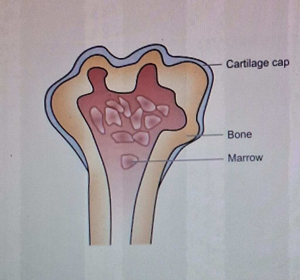
Benign tumour are generally spherical or ovoid in shape. They are encapsulated or well-circumscribed, more often firm, freely movable, and uniform, unless secondary changes like haemorrhage or infarction supervene.
Malignant tumours, on the other hand, are usually irregular in shape, poorly-circumscribed and extend into adjacent tissues. Secondary change like haemorrhage, infarction and ulceration are seen more often.
- Microscopic features
- Epithelial tumours
- Microscopic features
The epithelial tumours generally consist of acini, columns, sheets, or cords of epithelial tumour cells that may be arranged in solid or papillary pattern .
- Mesenchymal tumours
The mesenchymal tumours have mesenchymal tumour cells arranged as interlacing bundles, fasicles or whorls, lying separated from each other usually by intercellular matrix substance such as hyaline material in leiomyoma cartilaginous matrix in chondroma, osteoid in osteosarcoma, reticulin network in soft tissues sarcomas etc
- Local invasion (Direct spread)
- BENIGN TUMOUR.
Most benign tumours form encapsulated or circumscribed masses that expand and push aside surrounding normal tissues without actually invading, infiltrating or metastasising .
MALIGNANT TUMOUR
Malignant tumour also enlarge by expansion and some well-differentiated tumour may be
partially encapsulated as well e.g. follicular carcinoma thyroid. But characteristically, they are distinguished from benign tumour by invasion, infiltration and destruction of the surrounding tissue, besides distant metastasis.
Metastasis (Distant spread).
Metastasis (meta = transformation, stasis = residence) is defined as spread of tumour by invasion in such a way that discontinuous secondary tumour mass/masses are formed at site of lodgement.
Routes of Metastasis
Cancers may spread to distant sites by following pathways:
- Lymphatic spread
- Haematogenous spread
- Spread along body cavities and natural passages
(Transcoelomic spread, along epithelium-lined surfaces, spread via cerebrospinal fluid, implantation).
Effects of tumor on host
LOCAL EFFECTS
- Compression
- Mechanical obstruction
- Tissue destruction
- Infarction, ulceration, haemorrhage.
CANCER CACHEXIA
Patient with advanced and disseminated cancers terminally have asthenia (emaciation), and anorexia, together referred to as cancer cachexia (meaning wasting). Exact mechanism of cachexia is not clear but it does not occur due to the increased nutritional demands of the tumour.
FEVER.
Fever of unexplained origin may be presenting feature in some malignancies such as in Hodgkin’s disease, adenocarcinoma kidney, osteogenic sarcoma and many other tumours. The exact mechanism of tumour associated fever is not known but probably tumour cells themselves elaborate pyrogens.
TUMOUR LYSIS SYNDROME
This is a condition caused by extensive destruction of large number of rapidly proliferating tumour cells. The condition is seen more often in cases of lymphomas and leukaemias than solid tumour
and may be due to the large tumour burden (e.g. in Burkitt’s lymphoma), chemotherapy, administration of glucocorticoid or certain hormonal agents (e.g. tamoxifen).
PARANEOPLASTIC SYNDROMES.
Paraneoplastic syndromes (PNS) are group of conditions developing in patients with advanced cancer which are neither explained by direct and distant spread of the tumour, nor by the usual hormone elaboration by tissue of origin of the tumour. About 10 to 15% of the patients with advanced cancer develop one or more of the syndromes included in the PNS. Rarely, PNS may be the earliest manifestation of a latent cancer.
Pathological diagnosis of Tumors
- Histological Methods
These method are based on microscopic examination of properly fixed tissue (excised tumor mass or open/needle biopsy from the mass), supported with complete clinical and investigative data.
- Cytological Methods
Cytological methods for diagnosis consist of study of the cells shed off into body cavities (exfoliative cytology) and study of cells by putting a fine needle introduced under vacuum into the lesion (fine needle aspiration cytology, FNAC).
- Histochemistry and Cytochemistry
Histochemistry and cytochemistry are additional diagnostic tools which help pathologist in identifying chemical composition of cells, their constituents and their products by special staining methods.
- Immunohistochemistry
This is an immunological method of recognizing a cell by one or more of its specific components in cell membrane, cytoplasm or nucleus. These cell components (called antigens) combine with specific antibodies on formalin-fixed paraffin sections or cytological smears. The complex of antigen-antibody’s on slide is made visible for light microscopic identification by either fluorescent dyes (‘fluorochromes’) or by enzyme system (‘chromogens’).
- Electron Microscopy
Ultrastructural examination of tumor cell offers selective role in diagnostic pathology. EM examination may be helpful in confirming or substantiating a tumor diagnosis arrived at by light microscopy and immunohistochemistry.
- Tumour Markers (Biochemical Assays)
In order to distinguish from the preceding techniques of tumour diagnosis in which ‘stains’ are imparted on the tumour cells in section or smear, tumour markers are biochemical assays of products elaborated by tumour cells in blood or other body fluids. It is, therefore, pertinent to keep in mind that many of these products are produced by normal body cells too, and thus the biochemical estimation of product in blood or other fluid reflects the total substance and not by the tumour cells alone.