
10 Knee Ligament: Attachment & Function
Introduction
The knee is one of the strongest yet most vulnerable joints in our body—and its stability depends heavily on its knee ligament. Whether you’re walking, running, or simply standing, these strong bands of tissue keep everything in place and working smoothly. In this article, we’ll break down each knee ligament in a simple way—what it does, why it matters, and how injuries happen—so you understand your knee like never before. Let’s dive in!
The knee joint contains a total of 10 key knee ligaments when all major and provide knee support ones are included. Here’s the full list:
Primary (Main Stabilizing) Ligaments – 4
1. Anterior Cruciate Ligament (ACL)
2. Posterior Cruciate Ligament (PCL)
3. Medial Collateral Ligament (MCL)
4. Lateral Collateral Ligament (LCL)
Secondary (Supportive/Accessory) Ligaments – 6
5. Patellar Ligament
6. Oblique Popliteal Ligament
7. Arcuate Popliteal Ligament
8. Transverse Ligament of the Knee
9. Anterior Meniscofemoral Ligament (Ligament of Humphrey)
10. Posterior Meniscofemoral Ligament (Ligament of Wrisberg)
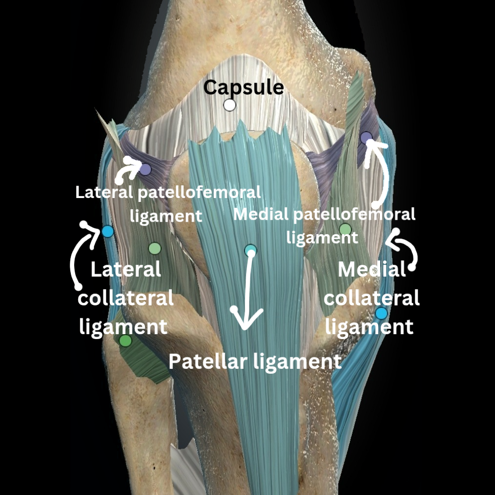
Medial Collateral Ligament (MCL)
The medial collateral ligament is made up of two distinct layers: a superficial and a deep portion, separated by a bursa.
- This knee ligament has a superficial layer which begins at the medial epicondyle of the femur and extends downward to attach on the upper medial part of the tibia.
- The deep layer of this knee ligament is the part of the joint capsule, originates from the lower portion of the medial femoral condyle and connects to the top of the medial tibial plateau.
Function of MCL of knee ligament
- It mainly prevents valgus stress (inward bending of the knee), especially when the knee is fully extended. The medial collateral ligament gets taut ( tighten up ) when the knee is in full extension.
- The MCL also assists in limiting forward movement of the tibia on the femur when ACL knee ligament function are compromised.
- With its rich blood supply, the MCL has good healing potential, so it often recovers naturally without surgery.
- In any circumstances if MCL got damaged , that time ACL ( Anterior cruciate ligament) get more strain. The majority of study says these circumstances increases the risk of injury to other knee ligaments.
Lateral Collateral Ligament (LCL)
The lateral collateral ligament (LCL) as the name suggests that it is located on the lateral side of the tibiofemoral joint. It originate from lateral femoral condyle goes to the head of the fibula where it joins with the tendon of the biceps femoris muscle to form the conjoined tendon. It is a extracapsular ligament which means outside of joint capsule.
Function of LCL of knee ligament
- The LCL helps the knee resist varus stress, or inward force that pushes the knee outward.
- It also helps control excessive outward rotation of the tibia.
Anterior Cruciate Ligament (ACL)
The Anterior cruciate ligament is a key knee ligament in the mid of the knee joint. It attaches to the front and lateral side of the tibial spine and extends upwards and backwards to connect to the medial side of the lateral femoral condyle.
It is made up of two different fiber bundles:
- Anteromedial bundle (AMB)
- Posterolateral bundle (PLB)
These fibers twist around each other during knee flexion.
Function of ACL of knee ligament
- The ACL is the primary stabilizer against forward movement of the tibia relative to the femur.
- It also prevents hyperextension of the knee.
- Additionally, it contributes to rotational stability, helping control twisting forces and preventing abnormal movement during side-to-side motions.
Posterior Cruciate Ligament (PCL)
The PCL connects from the back of the tibia, situated between the posterior horns of the menisci, and extends forward and upward to attach on the medial femoral condyle. Although it is within the joint capsule, it remains outside the synovial cavity.
The PCL also has two main bundles:
- Posteromedial bundle (PMB)
- Anterolateral bundle (ALB)
Function of PCL of knee ligament
It primarily resists backward movement (posterior translation) of the tibia, handling up to 90% of this load during most knee motions.
- The PCL helps to resist varus and valgus forces and helps control tibial rotation.
- Due to its structure, a backward force on the tibia may also result in slight external rotation, adding to its complex function.
Oblique Popliteal Ligament
The oblique popliteal ligament is a fibrous extension of the semimembranosus tendon that reinforces the posteromedial aspect of the knee joint capsule.It helps to stabilize the knee during full extension as it can extends diagonally across the back of the knee joint, and also work to resists hyperextension.
Posterior Oblique Ligament
Posterior oblique ligament is a knee ligament which is located deeper in the posterior knee.It provides additional support to the medial side of the knee joint. It originates just behind the adductor tubercle on the femur and attaches to the upper tibia, behind the attachment of the superficial medial collateral ligament. This knee ligament plays a major role in controlling valgus (inward bending) stress and contributes to posterior knee stability.
Arcuate ligament complex
The arcuate ligament is a Y-shaped thickening found in about 70% of knee. It reinforces the posterolateral corner of the knee joint capsule. This region also includes the lateral collateral ligament, iliotibial band, popliteus muscle and tendon, and the popliteofibular ligament. The arcuate ligament helps resist varus (outward bending) forces . This knee ligament helps to prevent excessive knee extension, especially when the knee is fully straightened.
Meniscofemoral Knee Ligaments
The meniscofemoral ligaments are specialized structures in the knee ligament system that vary in presence among individuals. Although not considered true ligaments—since they connect the lateral meniscus to bone rather than bone to bone—they play an important supportive role within the knee joint.
These knee ligaments originate from the posterior horn of the lateral meniscus and insert on the lateral aspect of the medial femoral condyle. There are two types:
- Ligament of Humphry, it is from PCL to anteriorly
- Ligament of Wrisberg, it is from PCl to posteriorly
Studies have shown that at least one of these knee ligaments is found in 91% of knees, and both may be present in about 30% of case.
Although the meniscofemoral ligaments are relatively small—having only around 14% of the cross-sectional area of the PCL. They can contribute significantly to knee stability.
Function of these knee ligament
- They assist the posterior cruciate ligament in limiting posterior translation of the tibia
- They may also help the popliteus muscle in controlling lateral rotation of the tibia.
- This knee ligament helps in knee joint integrity and functional movement.