
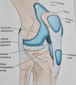
Lateral
- A bursa deep to lateral head of gastrocnemius.
- A bursa between fibular collateral ligament and the biceps femoris.
- A bursa between the fibular collateral ligament and the tendon of the popliteus.
- A ursa between the tendon of the popliteus and the lateral condyle of the tibia
Medial
- A bursa deep to medial head of the gastrocnemius.
- The anserine bursa is a complicated bursa which separates tendons of the Sartorius , the gracilis and the semitendinosus from one another , from tibia , and from the tibisl collateral ligament.
- A bursa deep to tibial collateral ligament
- A bursa deep to semimembranosus.
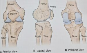
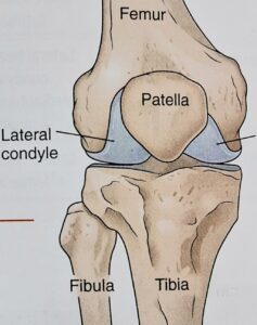
Relations of knee joint
ANTERIORLY
Anterior bursae , ligamentum patellae and patellar plexus of nerves.
POSTERIORLY
- At the middle; popliteal vessel , tibial nerve.
- Posterolaterally ; lateral head of gastrocnemius, plantaris and common peroneal nerve .
- Posteromedially; medial head of gastrocnemius semitendinosus, semimembranosus, gracilis , and popliteus at its insertion.
- Sartorius , gracilis and semitendinosus
- Great saphenous vein with saphenous nerve.
- Semimembranosus.
- Five genicular branches of the popliteal artery.
- The descending genicular branch of femoral artery
- The descending branches of the lateral circumflex femoral artery
- Two recurrent branches of the anterior tibial artery .
- The circumflex fibular branch of posterior tibial artery .
- Femoral nerve , through it is branches to vasti, especially vastus medialis .
- Sciatic nerve , through thye genicular branches of the tibial and common peroneal nerve
- Obturator nerve , through it's posterior division .
- Fibrous capsule
- Ligamentum patellae
- Tibial collateral or medial ligament
- Fibular collateral or lateral ligament
- Oblique popliteal ligament
- Arcuate popliteal ligament
- Anterior cruciate ligament
- Posterior cruciate ligament
- Medial meniscus
- Lateral meniscus
- Transverse ligament
- Anteriorly , it is deficient
- Posteriorly ,it is attached to the intercondylar line
- Posteriorly ,it is attached to the intercondylar line.
- Anteriorly , it descends along the margins of the condyles to the tibial tuberosity , where it is deficient .
- Posteriorly , it is attached to the intercondylar ridge which limits the attachment of the posterior cruciate ligament .
- Posterolaterally , there is a gap behind the lateral condyle for passange of tendon of popliteus.
- The transverse axis around which these movements take place is not fixed. During extension, the axis moves forwards and upwards, and in the reverse direction during flexion. The movement are invariably accompanied by rotations or conjuct rotation. When the foot is on ground , while standing erect , medial rotation of femur occurs during last 30 degree of extension as in position of “ attention “ by the vastus medialis. It is called conjunct rotation. During position of “stand at ease “. There is lateral rotation of femur, during initial stages of flexion, by popliteus muscle
MEDIALLY
LATERALLY
Biceps femoris and tendon of origin of popliteus .
Blood supply
The knee joint is supplied by anastomoses around it , the chief source of blood supply are:
Nerve supply
Ligament
The knee joint is supported by the following knee joint ligament
Fibrous capsule
The fibrous capsule of knee joint is very thin and is deficient anteriorly, where it is replaced by the quadriceps femoris, the patella and the ligamentum patellae.
Femoral attachment
It is attached about half to one centimeter beyond the articular margins. The attachment has three special features.
Tibial attachment
It is attached about half to one centimeter beyond the articular margins. The attachment has three special feature
Movement
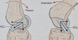
Active movement at the knee are flexion, extension, medial rotation and lateral rotation.
Flexion and extension are the chief movement. These take place in the upper compartment of the joint, above the menisci. They differ from the ordinary hinge movement in two ways.
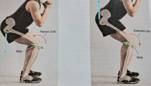
Medial rotation of the femur occurs during the last 30 degree of extension, and lateral rotation of the femur occurs during the initial stages of flexion.
What is the best diet to lower LDL cholesterol?
What is the best diet to lower LDL cholesterol? Introduction If you are concerned about high cholesterol, especially elevated LDL (Low-Density Lipoprotein) cholesterol, making the
What is best diet for high cholesterol ?
What is best diet for high cholesterol ? Maintaining healthy cholesterol levels is essential for protecting the heart and reducing the risk of cardiovascular
Best 25 Heart Healthy Foods to Eat
Best 25 Heart Healthy Foods to Eat Heart disease remains one of the leading health concerns worldwide, but many risk factors can be managed through
Sharp Pain in the Heel of My Foot: Causes and Treatment
Sharp Pain in the Heel of My Foot: Causes and Treatment Pain in the heel of my foot is one of the most common foot
Why my heel hurts when i walk : Possible Reasons and Fixes
Why my heel hurts when i walk : Possible Reasons and Fixes Heel pain can make everyday activities difficult. Many people notice discomfort while getting

8 best pain killers for muscle pain
8 best pain killers for muscle pain Pain is one of the most common health complaints worldwide, and pain killers are often the first solution