
Ligament Of Elbow joint
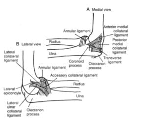
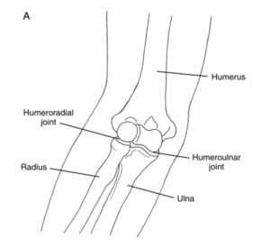
- Capsular ligament ( Medial collateral ligament ): superiorly, the attached to the lower end of the humerus in such a way that the capitulum the trochlear, the radial fossa, the coronoid fossa, and the olecranon fossa are intracapsular. Inferomedially, it is attached to the margin of the margin of the trochlear notch of the ulna except laterally; inferolateral; it is attached to the annular ligament of the superior radioulnar joint. The synovial membrane lines capsule and fossae.
The anterior ligament and posterior ligament are thickening of the capsule. - Ulnar collateral ligament: In the elbow joint, this ligament is triangula. Its apex is attached to the medial epicondyle of the humerus, and its base to ulna. The ligament has thick anterior and posterior bands: These are attached below to the coronoid process and olecranon process, respectively of the elbow joint. Their lower ends are joined to each other by an oblique band which gives attachment to the thinner intermediate fibers of the ligament. The ligament is crossed by the ulnar nerve and it gives origin to the flexor digitorum superficialis. It is closely related to the flexor carpi ulnaris and the triceps brachii.
- Capsular ligament ( Medial collateral ligament ): superiorly, the attached to the lower end of the humerus in such a way that the capitulum the trochlear, the radial fossa, the coronoid fossa, and the olecranon fossa are intracapsular. Inferomedially, it is attached to the margin of the margin of the trochlear notch of the ulna except laterally; inferolateral; it is attached to the annular ligament of the superior radioulnar joint. The synovial membrane lines capsule and fossae.
The Function of Elbow joint.
It provides lateral support to the elbow joint.
It helps to resist varus stress in the elbow joint.
It provides a dynamic stabilizer of the elbow joint.
A radial collateral ligament is a fan-shaped band extending from the lateral epicondyle to the annular ligament. Its gives origin to the supinator and to the extensor carpi radialis brevis.
Function
- Reinforcement for humeroradial articulation
- It resists varus stress.
- Resistance to longitudinal traction.
- Works as primary soft tissue restraint.
Relation
- Anteriorly: Brachialis, median nerve, brachial artery, and the tendon of biceps brachii
- Posteriorly: Triceps brachii and anconeus.
- Medially: Ulnar nerve, flexor carpi ulnaris, and common flexor.
- Laterally: Supinators, extensor carpi radialis bervis, and other common extensors.
Blood supply is from anastomoses around the elbow joint.
Nerve Supply of Elbow joint
The joint receives branches from following nerves.
- Median nerve
- Ulnar nerve
- Radial nerve
- Musculocutaneous nerve through its branch to the brachialis
Movement Of Elbow joint.
- Flexion is bought about by:
- Brachialis.
- Biceps brachii.
- Brachioradialis.
- An extension is produced by:
- Triceps brachii.
- Anconeus.