
Spinal Cord
Table of Contents
What is Spinal cord ?
The spinal Cord is a long tube that extends from the foramen magnum where it is continuous with medulla oblongata, above and up to the lower border of the first lumbar vertebra below. It is lies loosely in the vertebral canal.
Coverings of Spinal cord :
Spinal cord is covered by the sheaths called meninges, which are the membranous in nature. Meninge are dura mater, pia mater and arachnoid mater. Meninges are responsible for the protection and nourishment of the nervous tissues.
Shape and Length of Spinal cord:
The Spinal cord is cylindrical. The length of the spinal cord is about 43 cm in females and about 45 cm in males
31 Segments of Spinal cord :
Spinal cord is made up of 31 segments. Segments of the spinal cord correspond to 31 pairs of spinal nerves symmetrically.
1. Cervical segments/Cervical spinal nerves = 8
2. Thoracic segments/Thoracic spinal nerves = 12
3. Lumbar segments/Lumbar spinal nerves = 5
4. Sacral segments/Sacral spinal nerves = 5
5. Coccygeal segment/Coccygeal spinal nerves = 1
Total = 31


Conus Medullaris and Filum Terminale
Below lumbar enlargement, the spinal cord rapidly narrows to a cone-shaped termination called conus medullaris. A slender non-nervous filament called filum terminale extends from conus medullaris downward to the fundus of the dural sac at the level of the second sacral vertebra.
Gray matter of spinal cord
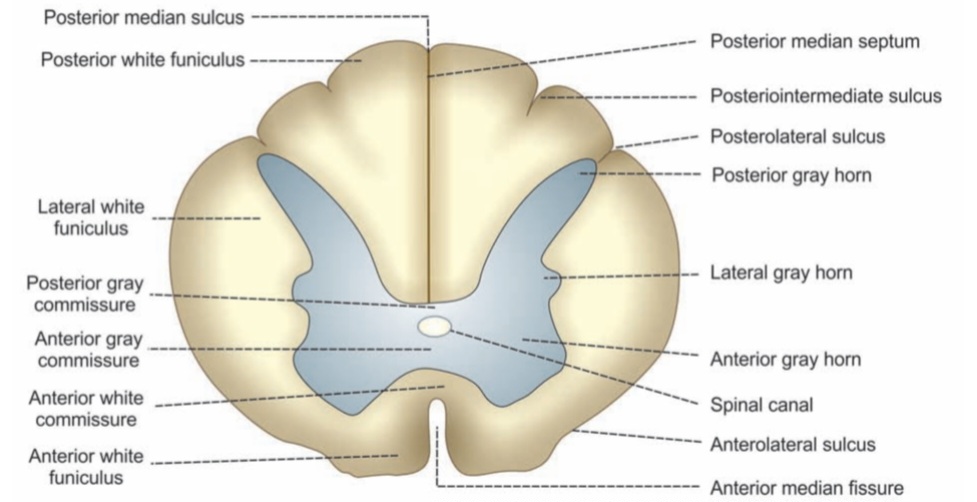
The gray matter of spinal cord is the collection of nerve cell bodies, dendrites, and parts of axons. Its placed centrally in form of wings of the butterfly and it resembles the letter ‘H’. Exactly in the center of gray matter, there is a canal called the spinal canal.
- Ventral and dorsal portions of each lateral half of gray matter are called ventral (anterior) and dorsal (posterior) gray horns respectively.
- The gray matter forms a small projection in between anterior and posterior horns in all thoracic and first two lumbar segments. It is called lateral gray horn.
- Part of the gray matter anterior to central canal is called anterior gray commissure
- Part of gray matter posterior to central canal is called posterior gray commissure.
Neurons in Gray Matter of Spinal Cord
1. Golgi type I neurons
Golgi type I neurons have a long axons and are usually found in the anterior horns. Axons of these neurons form long tracts of spinal cord.
2. Golgi type II neurons
Golgi type II neurons have short axons, which are found mostly in posterior horns of spinal cord. Axons of these neurons pass towards the anterior horn of the same side or opposite side.
How these neurons are organized?
The organization of neurons in the gray matter of the spinal cord is described in two methods:
1. Nuclei or columns
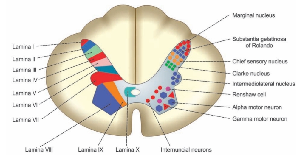
Nuclei in Posterior Gray Horn of spinal cord
- Marginal nucleus
- Substantia gelatinase of Rolando
- Chief sensory nucleus or nucleus proprius
- Dorsal nucleus of Clarke
Nuclei in Anterior Gray Horn of spinal cord
- Alpha motor neurons
- Gamma motor neurons
- Renshaw cells
2. Laminae or layers
Laminae in Posterior Gray Horn
Laminae I to VI constitute posterior gray horn. These laminae contain nuclei of sensory neurons, which are concerned with the sensory functions of body.
Lamina in Lateral Gray Horn
Lateral gray horn contain only one lamina, the lamina VII. It contains intermediolateral nucleus of spinal cord.
Laminae in Anterior Gray Horn
Laminae VIII and IX form the anterior gray horn. These laminae contain nuclei of motor neurons, which are concerned with motor functions of body .


Fissure and Sulci
Anterior median fissure
The Anterior surface of the spinal cord, there is a deep furrow known as anterior median fissure. Depth of this fissure is about 3mm.
Anterolateral sulcus
Lateral to the anterior median fissure on either side, there is a slight depression in spinal cord called the anterolateral sulcus.
Posterior median sulcus
On the posterior aspect, there is a depression in spinal cord called posterior median sulcus
Posterior median septum
Posterior median sulcus. This sulcus is continuous with a thin glial partition called posterior median septum.
Posterior intermediate sulcus
Lateral to posterior median sulcus, there is a posterior intermediate sulcus. It is continuous with posterior intermediate septum, which extends for about 3 mm into spinal cord.
Posterolateral sulcus
Lateral to posterior intermediate sulcus, is the posterolateral sulcus.
White matter of spinal cord
White matter of spinal cord surrounds the gray matter. It is the formed by bundles of both myelinated and non-myelinated fibers, but predominantly myelinated fibers. The band of white matter lying in the front of anterior gray commissure is called anterior white commissure
White matter of spinal cord into three white columns .
I. Anterior or Ventral White Column
Ventral white column lies between anterior median fissure on one side and anterior nerve root and anterior gray horn on the other side. Its also called anterior or ventral funiculus.
II. Lateral White Column
Lateral white column is present between anterior nerve root and anterior gray horn on one side and posterior nerve root and posterior gray horn on the other side. It is also c/a lateral funiculus.
III. Posterior or Dorsal White Column
Dorsal white column is situated between posterior nerve root and posterior gray horn on one side and posterior median septum on other side. It is also called posterior or dorsal funiculus.
Spinal cord injury and incidence
Globally, it is estimated that about three million people are living with spinal cord injuries, and in the United States alone, nearly 12,000 new cases are reported each year.
In the United Kingdom, the annual rate of acute spinal cord injuries (SCI) is around 19 new cases per million individuals, with approximately 50,000 people currently living with the condition. The majority of these injuries result from traumatic events, most commonly due to road traffic accidents and falls.
We assess spinal cord injuries with the help of American Spinal Injury Association (ASIA) International Standards for Neurological Classification of Spinal Cord Injury (ISNCSCI), which is a globally accepted tool for evaluating and describing such injuries.
What are the functions of spinal cord ?
The spinal cord functions as an intricate communication network that transmits signals between the brain and the rest of the body. It consists of several specialized anatomical pathways, each responsible for carrying specific types of sensory and motor information.
The spinal cord serves as the primary processing center for pain signals before they are transmitted to the brain. Sensory nerve fibres deliver messages from the body to the dorsal horn of the spinal cord, where they interact with other neurons. These spinal neurons then relay the information to higher brain centers, enabling the perception of both normal sensations and pain.
The spinal cord regulates these incoming signals through various control systems, such as excitatory and inhibitory neurons, NMDA receptor activity, and descending inputs from the brainstem that can either amplify or suppress pain. When an injury or inflammation occurs, these control systems undergo changes that heighten the spinal cord’s responsiveness to sensory input. This excessive sensitivity, known as central sensitization, contributes to the persistence of chronic pain.
In such conditions, the normal balance between pain-enhancing (excitatory) and pain-inhibiting (inhibitory) neurons becomes disrupted. As a result:
Excitatory activity rises, intensifying pain transmission.
Inhibitory control weakens, reducing the spinal cord’s ability to suppress pain signals.
Consequently, neurons in the dorsal horn become overactive and send amplified pain messages to the brain, reinforcing the cycle of central sensitization and chronic pain.

