
X-Ray Imaging
Table of Contents
X-ray imaging is a diagnostic medical procedure used in various clinical conditions such as fracture, Decalcification, Infection, and Tumors. In X-ray Imaging technology there is an use of X-ray. These are electromagnetic radiation, similar to light rays but it has high energy.
Patient preparation before X-ray imaging?
- All radio-opaque/metal items should be removed from the area of examination eg, including hair clip, pins, belts, etc
- Ponytail /Bunches of hair often produce artifacts and thus should loosen.
- If the area of interest includes the mouth then false teeth or metal dental bridge should be removed unless the patient has injuries.
- The patient should clean or bath before X ray
- If there is an X-ray of the abdomen then the patient needs to be empty bowels.
Types of X Rays
According to the view of x -ray taken there are three types of X-ray .
1 . Anterior view
2. Posterior view
3. Lateral view
Anterior-posterior view X-Ray
This is a projection of the image from the anterior to the posterior side. The X-rays passes from anterior to posterior side of the human body.
Position of the patient &Image receptor
- The patient is asked to lie down in a supine position on the X-ray table or erect against the becky detector in a true anterior-posterior position.
- The median sagittal plan of the body is adjusted & brought at a right angle to the image detector.
Posterior view X-ray
This is a projection of the image from posterior to anterior side. The X-rays passes from posterior to anterior side of human body.
Position of patient & Image receptor
- The patient lies down on the imaging table in a prone position or erect facing towards the Becky detected with the median sagittal plan at the right angle & co-incident with the midline of the Becky.
- The patient fashed & the nose should be touching the becky detector perpendicular to the image detectors.
- In skull X-ray, the defects should be 2 inches above the base of the skull. Ensure that the mid-part of the frontal bone is positioned in the center of the Becky.
Lateral view x-ray
This view is also known as Schuller’s view . It is principally used for viewing mastoid cells which are present lateral side of the skull.
Position of patient & Image receptor
- Ask a patient to lie down on the X-ray table in a prone position erect facing towards the becky detects.
- In skull X-ray, the chin should be elevated so that the head is in a lateral position and the median sagittal plan is parallel to the becky.
- The casette’s upper edge is kept 2 inches above the base of the skull
Chest X-ray for lung disease
The ‘plain’ chest X-ray is performed on the majority of patients suspected of having chest disease. A posteroanterior (PA) film provides information on the lung fields, heart, mediastinum, vascular structures, and thoracic cage.

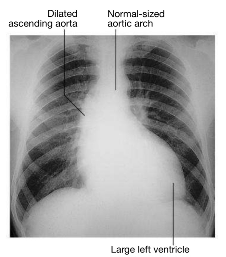
Chest X-ray for Heart disease
Chest X-ray is useful for determining the size and shape of heart, and the state of pulmonary blood vessels and lung fields. Most information is given by postero-anterior (PA) projection taken in full inspiration. Anteroposterior (AP) projections can be performed when the patient movement is restricted but results in magnification of the cardiac shadow.
Dual X-ray absorptiometry for rheumatic condition
Dual X-ray absorptiometry can estimation of bone mineral density (BMD) is used for diagnosis and management of osteoporosis. Measurements at the lumbar spine, hip, and sometimes forearm are obtained.
Example: Osteoporosis is defined in postmenopausal women and men of more than 50 years old with a T-score of 2.5 or below (shaded red in the figure). BMD values above −1.0 and below +2.5 are considered normal (yellow/green), whereas values above +2.5 indicate high bone mass, the most common cause being OA. The results need to be interpreted carefully and in reference to coexisting conditions, such as aortic calcification, vertebral fractures, degenerative disc disease and OA, all of which can artefactually raise BMD results. Radiographic correlation is then advisable.
- X-rays by the National Institute of Biomedical Imaging and Bioengineering