
Ultrasound Therapy
Table of Contents
What is Ultrasound?
A therapeutic Ultrasound that produces a high-frequency sound wave greater than 20,000 Hz is called High-Intensity Focused Ultrasound (HIFU). The frequencies used for therapeutic purposes range mainly from 1 MHz to 3 MHz and are used in physical therapies for various clinical conditions.
Therapeutic Ultrasound is different from diagnostic ultrasound based on its uses in Hospitals. Diagnostic ultrasound is used to diagnose various conditions by taking images of internal organs, whereas Therapeutic ultrasound is commonly used by physical therapists to treat pain and promote healing.
The major condition where Ultrasound is used
- Musculoskeletal conditions: myofascial pain, frozen shoulder, intense shoulder pain, Osteoarthritis.
- Tissue injury: swelling of muscles, joints, and ligaments, delayed wound healing, Muscle spasms.
- Scar tissue
- Cancer
The Ultrasound is made as such that for a 1MHz machine a vibrating source with a frequency of one million cycles per second is needed . This is achieved using either a quartz or a barium titanite crystal. These crystal deform when subjected to a varying potential difference - a piezo - electric effect.Components of Ultrasound
- Source of high-frequency current
- Co-axial cable: It helps to convey the current to the transducer
- Transducer head
- Link electrode
- Quart crystal Current passes to Quartz and changes shape.
- Metal front plate: It causes the movement of the metal front plate which produces Ultrasonic waves.
As soon as the Ultrasound head is placed directly on the skin then the waves originating from the source undergo reflection leading to overheating. let’s discuss this in detail.
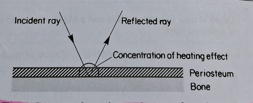
Reflection
- Sound wave obeys the laws of reflection and if an ultrasonic beam traveling through one medium encounters another medium which will not transmit it
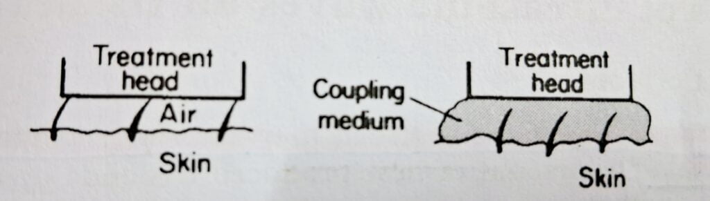
- Coupling medium is used to exclude air from space between treatment head and skin .
- In periosteum region , Reflection from bone double the intensity of ultrasound lead to over heating . It lead to Periosteal pain .
When the waves enter the skin through any coupling media giving a Medium other than air helps in the refraction of these waves Let’s understand in brief.
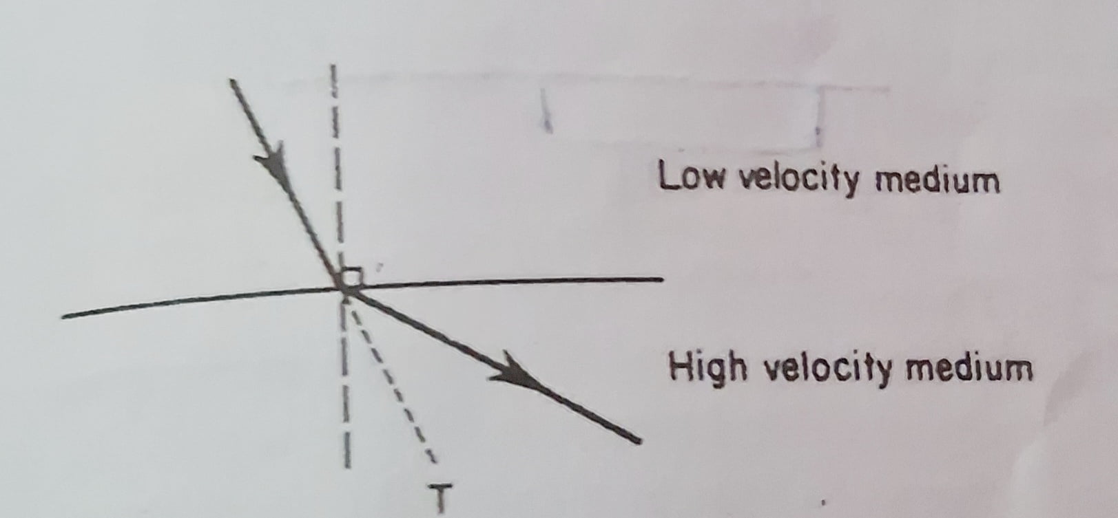
Refraction
- A sound wave is perpendicular to the target tissue to have less refraction.
- When traveling from a medium in which its velocity is low into one in which its velocity is high, it is refracted away from the normal .
Standing waves are produced leading to inhibition of blood flow. It causes the blood cells together. These are prevented by continuous concentric movement of head of ultrasound
Coupling media
- Coupling medium is used to exclude air from space between treatment head and skin .
- Example : Aqua sonic gel have 72.6 % and 67% of transmission
Attenuation
- Ultrasound penetrates skin depth and is absorbed by muscle converting to heat and less energy.
- It is due to the absorption and scattering of waves.
- Cartilage, tendon, ligament, and blood vessels contain proteins that can absorb more
- Mode
- Pulsed mode: In this mode, there is intermittent transfer of sound waves. It has mechanical effects on the body.
- Continuous mode: In this mode, there is a continuous transfer of sound waves . It has a thermal effect on the body
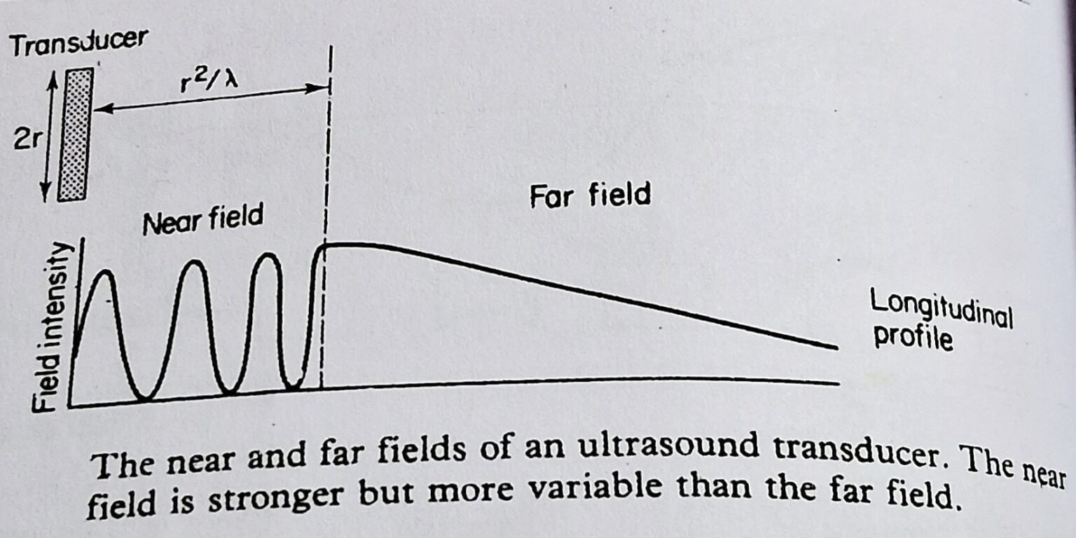
Depth of near flown from 1MHz and 3Mhz ultrasonic transducers of different sizes.
1 MHz transmits waves superficially, used for acute conditions.
3 MHz transmit waves in deep, used for chronic conditions.
| Transducer radius(mm) | Frequency (MHz) | Extend of near field ( cm ) |
| 15 | 1 | 15 |
| 15 | 3 | 45 |
| 10 | 1 | 6.5 |
| 20 | 1 | 26.5 |
Effects of ultrasound on tissue
Thermal effects
Pulsed ultrasound has little thermal effect, whereas continuous ultrasound mode give more of thermal effect which help to reduce swelling and chronic condition .
Mechanical effects
The Principal in which the mechanical effect of ultrasound work is cavitation is a condition in which a bubble of gases is produced in tissue. There are two different types of cavitation produced in tissues.
Stable cavitation is not dangerous to tissue. Bubbles remain intact and oscillate harmlessly.
Transient cavitation is dangerous to tissue and bubbles grow and collapse rapidly.
Cavitation is reduced by some factors such as :
- Using intensity below 4 watts/ cm square
- Using pulsed source.
- Moving treatment head in circulation motion continuously.
Chemical & biological effects
- Acoustic streaming is a unidirectional flow of tissue components that occurs in the cell membrane.
- It will wash out the ion.
- The permeability of the membrane increases.
- Ions entry increases.
- Messenger activity increases in cells.
- Pain relief by pain gate theory
Indication
Indication of Ultrasound theraphy indicate the various condition in which we can apply ultrasound , such as
Recent injury
- Remove traumatic exudate.
- Reduce adhesion formation.
- Promote the rate of repair of damaged tissue.
- delayed wound healing
Inflammation
- swelling of muscles, joints, and ligaments,
- Muscle spasms.
- Scar tissue
- Chronic indurated edema ( by breakdown of adhesion )
Application of Ultrasound Therapy
When any patient undergo a full assessment program by any therapist and a diagnosis of the condition is completed then the treatment starts, if treatment includes an ultrasound (for example, if any person has swelling ) then we apply ultrasound
Steps for application of ultrasound therapy
- Collect every material you need during the treatment such as ( cotton or tissue , gel)
- Then plugin the main switch and ON from electric board.
- Take the gel and apply on treatment head and place the head perpendicular to the targeted area.
- Ultrasound machine have separate ON /OFF button then ON the machine .
- Set frequency and time
- Increase the intensity slowly and wisely according to the tolerance of patient.
- During treatment move the head continously without stoping for sec0nd .
- Then slowly reduce the intensity and then switch OFF the machine .
- You can even Plug off the wire .
The ultrasound is applied in three ways explained by
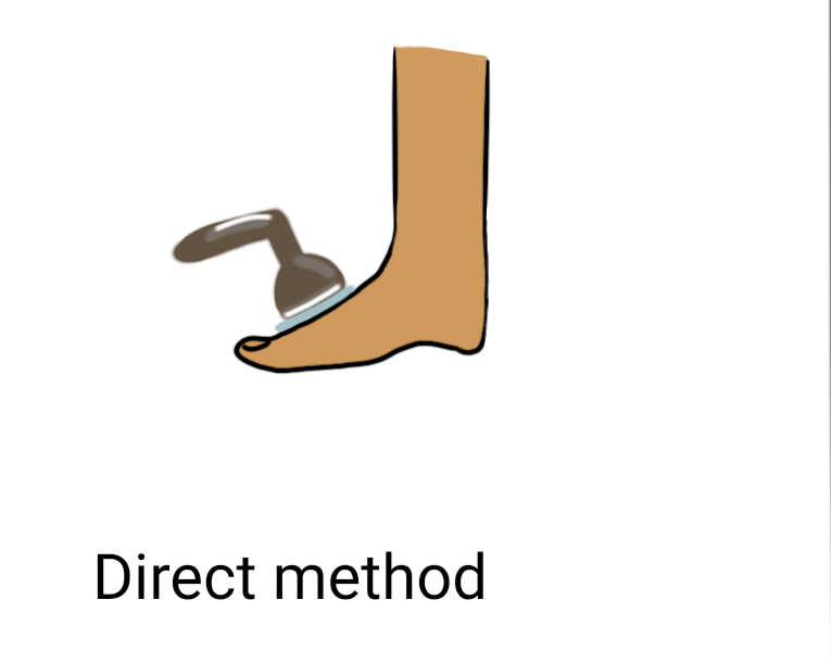
Direct contact Method
- The ultrasound head is in direct contact with the skin where a coupling medium is used between them.
- The head moves in a concentric circle to avoid concentration at one point.
- The ultrasound remains in contact during the turned-on and turned-off machine.
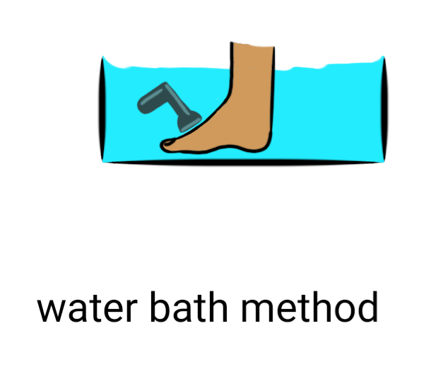
Water bath Method
- Water filled with de-gassed water is used between the head and skin.
- Tap water is avoided to uses as it forms gas bubbles that can lead to cavitation.
- The head should be 1cm from the skin.
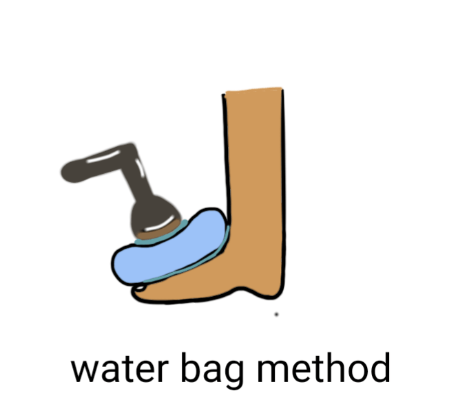
Water bag Method
- A rubber bag with de-gassed water is used between the head and skin.
- A coupling medium is used between the rubber bag and skin and also between the rubber bag and treatment head.
Acute condition
Initial stage
- 0.25 or 0.5 watt / cm 2
- 2 -3 min
- pulsed beam
- If condition improve , same dose is repeated .
Later stage
- 0.8 watt / cm 2
- 4-5 min
Sometimes symptoms may indicate that repair process taking place .
Chronic condition
we can use both pulsed and continous mode.
Intial Stage
- 0.8watt/cm 2
- 4 min
If beneficial effect is visible then repeted next time.
History of Ultrasound
The Ultrasound first documented and published in 1958 by Ian Donald in Glasgow which was first utilized sonography as a diagnostic tool used in obstetrics and gynecology.With technological advancements have allowed ultrasound to become a therapeutic modality which is used to treat various musculoskeletal condition like osteoarthritis, and myofascial pain etc.
Contraindication
Contraindication of ultrasound explains that various situation in which the ultrasound usage is dangeours .To avoid danger every therapist should know when to use and when we cann0t use .
- Over the uterus during pregnancy
- Over the gonads
- Over malignancies and precancerous lesions
- On patients with vascular abnormalities, i.e.deep vein thrombosis, emboli, severe
atherosclerosis - Over the eye
- Over the stellate ganglion
- For hemophiliacs not covered by factor
replacement - Over the spinal cord after laminectomy
- Directly over metal implants
- Over an electronic device
- Over tissues previously treated with deep Xray or radiation
- Tuberculosis (local)
- Over damaged or at risk skin, i.e. skin rash,eczema
- Over anesthetic areas
- Over excitable tissue, i.e. heart, exposed
nerve, carotid sinus
Some patient with certain condition explains where we cannot use ultrasound such as
Vascular condition
Conditions such as thrombophlebitis , where insonation may cause emboli to be broken off, are not treated with ultrasound .
Acute sepsis
An area which presents acute sepsis is not treated with ultrasound because of the danger of spreading the inflection .
Radiotherapy
Radiotherapy has an adverse effect on the tissue , therefore ultrasound is not applied to a radiated area for at least six month after irradiation
Tumors
Tumor are not insonated because they may be stimulated into growth or throw off metastases
Pregnancy
A pregnant uterus is not treated as the insonation might produce fetal damage ( Ultrasonic scanning as a diagnostic aid in pregnancy is different from that used for therapeutic purpose)
Cardiac disease
- Patients who have had cardiac treaded with low intensities in order to avoid sudden pain and area such as the cervical ganglion and the vague nerve are avoided because of the risk of cardiac stimulation .
- Patient fitted with cardiac pacemaker are not usually treated with ultrasound in the area of the chest , as the ultrasound generator may have an effect on the pacemaker’s rate of stimulation.
Danger
Danger indicate that the various unusual dangerous situation which can cause due to carelessness of the user before , during and after treatment of patient .These can be
- Burns
- Cavitation
- Overdose
- Damage to equipment
- Electric shock
Recent Article post
Recent Article
How to Cure Insomnia Naturally ?
How to Cure Insomnia Naturally If you’re struggling with insomnia, the good news is that effective treatment doesn’t always require medication. Research shows that several
Understanding your back problems at once
Understanding your back Problems at once Introduction Because you clicked on this article, it is likely that you have had firsthand experience of how frustrating
What is the best diet to lower LDL cholesterol?
What is the best diet to lower LDL cholesterol? Introduction If you are concerned about high cholesterol, especially elevated LDL (Low-Density Lipoprotein) cholesterol, making the
What is best diet for high cholesterol ?
What is best diet for high cholesterol ? Maintaining healthy cholesterol levels is essential for protecting the heart and reducing the risk of cardiovascular

Best 25 Heart Healthy Foods to Eat
Best 25 Heart Healthy Foods to Eat Heart disease remains one of the leading health concerns worldwide, but many risk factors can be managed through
Sharp Pain in the Heel of My Foot: Causes and Treatment
Sharp Pain in the Heel of My Foot: Causes and Treatment Pain in the heel of my foot is one of the most common foot