


What is the best diet to lower LDL cholesterol?
What is the best diet to lower LDL cholesterol? Introduction If you are concerned about high cholesterol, especially elevated LDL (Low-Density Lipoprotein) cholesterol, making the
What is best diet for high cholesterol ?
What is best diet for high cholesterol ? Maintaining healthy cholesterol levels is essential for protecting the heart and reducing the risk of cardiovascular
Best 25 Heart Healthy Foods to Eat
Best 25 Heart Healthy Foods to Eat Heart disease remains one of the leading health concerns worldwide, but many risk factors can be managed through
Sharp Pain in the Heel of My Foot: Causes and Treatment
Sharp Pain in the Heel of My Foot: Causes and Treatment Pain in the heel of my foot is one of the most common foot
Why my heel hurts when i walk : Possible Reasons and Fixes
Why my heel hurts when i walk : Possible Reasons and Fixes Heel pain can make everyday activities difficult. Many people notice discomfort while getting
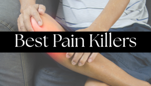
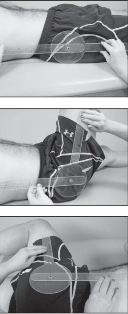
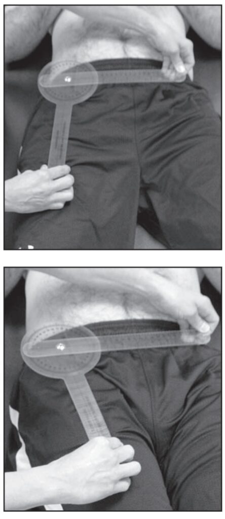
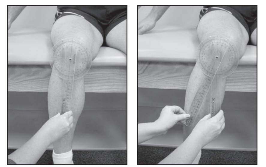
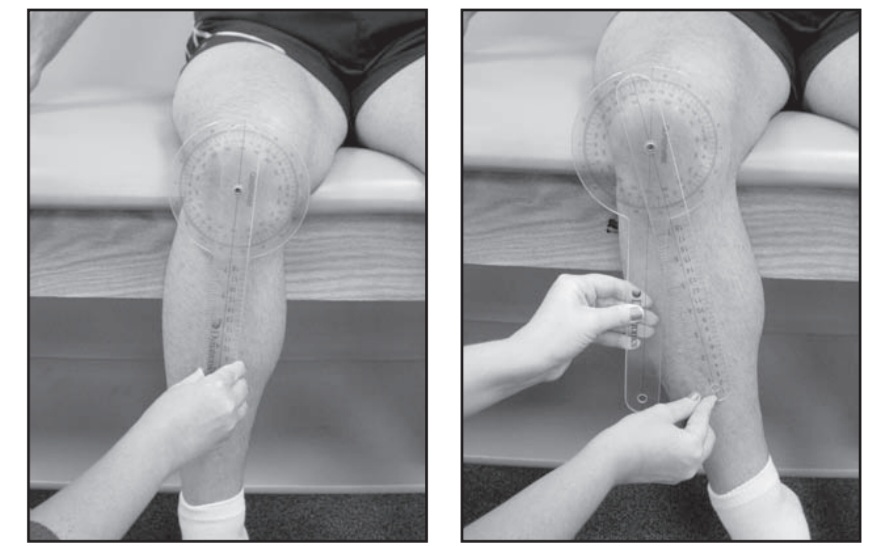
8 best pain killers for muscle pain
8 best pain killers for muscle pain Pain is one of the most common health complaints worldwide, and pain killers are often the first solution