
Tetralogy of Fallot
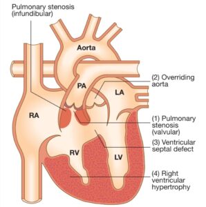
Tetralogy of Fallot is a congenital cardiac malformation or abnormalities that consists of :
2. Obstruction of the right ventricular outflow tract,
3. Override of the ventricular septum by the aortic root,
4. Right ventricular hypertrophy.
This most common in children born with cyanotic heart disease.
Cyanosis( bluish color in the skin, lips, and nail beds) may vary from mild to severe, and patients may present as newborns or, more commonly, young infants. Infants with classic tetralogy of Fallot and stable anatomy should undergo or go through primary complete intracardiac repair. The overall hospital mortality is approximately 3 to 5 percent, with most patients who survive having an excellent clinical and hemodynamic result.
Pathophysiology
The human heart development starts around the 20th day of gestation, with the fusion of the outer endocardial tubes into a single tubular structure( cardiac tube). Subsequently, cardiac tube folds and loops, with development of an atrium that is cranial and dorsal, and a primitive ventricle is moving downward, ventrally, and to the right. The dominant chamber of heart, the right ventricle of the embryo and fetus, receives 65% of the venous return and is the main contributor to the lower part of the body, the placenta, and the lungs. The embryologic process that contributes to the development of the tetralogy of Fallot still is unknown. Still, an association that had been observed is an anterior and cephalad deviation of the infundibular septum that results in a misaligned ventricular septal defect, with an overriding aortic root causing a subsequent right ventricular outflow obstruction.
The patient with tetralogy of Fallot shows ventricular septal defects usually peri membranous that can extend into the muscular septum. Different factors can contribute to right ventricular outflow obstruction, including pulmonary valve that is usually bicuspid and stenotic, the hypoplastic pulmonary valve annulus, the deviation of infundibular septum that causes a subvalvular obstruction, and the hypertrophy of muscular bands in this region. The degree of overriding aorta usually varies and receives blood flow from both ventricles. The physiological process surrounding the hypercyanotic episodes or “Tet spells” in tetralogy of Fallot consists of either a decrease in systemic vascular resistance or increase in pulmonary resistance contributing to a right-to-left shunt across ventricular septal defect, causing marked desaturation.
What is the cause of tetralogy of Fallot?
The etiology is multifactorial, but majorly caused by the following factors:
1. Untreated maternal diabetes,
2. Phenylketonuria,
3. Intake of retinoic acid
4. Associated chromosomal anomalies can include trisomies 21, 18.
5. Microdeletions of chromosome 22.
In most children, cause of tetralogy of Fallot isn’t known. It’s a common type of heart defect. It may be seen more commonly in children with Down syndrome or DiGeorge syndrome.
Sign and Symptom
Symptoms may include:
- The blue or gray coloration of the skin.
- Shortness of breath and rapid breathing, especially during activities or exercise.
- Trouble gaining weight.
- Weakness
- Irritability.
- Crying for long periods.
- Fainting.
When to see a doctor?
Congenital heart defects are often diagnosed before or soon after your child is born when you notice the following symptoms kindly seek medical help.
- Trouble breathing.
- More irritable than usual.
- Bluish color of the skin.
- Seizures.
- Weakness
How to diagnose Tetralogy of Fallot?
Tetralogy of Fallot can be diagnose by chest radiograph, electrocardiogram, and echocardiogram. The echocardiogram establishes the definitive diagnosis and can provide sufficient information for surgical treatment planning.
Chest radiographs
Chest radiographs usually show normal-size heart silhouette, with an upturned apex and a concave main pulmonary artery segment, commonly known as “boot-shaped”.
Electrocardiogram
Its common to see signs of right atrial enlargement and right ventricular hypertrophy showing right axis deviation, prominent R wave anteriorly and S waves posteriorly, upright T wave in V1 (abnormal after 7 days of life up to 10 years of age), and QR pattern in the right precordial leads.
Echocardiogram
An echocardiogram is a gold standard, addressing the anatomy and severity of the right ventricular outflow obstruction, the location and number of ventricular septal defects, and assessing associated anomalies or variants with the coronaries arteries and aortic arch.
What is the treatment of Tetralogy of Fallot?
Tetralogy of Fallot is a congenital defect in the heart that needs surgery to fix the heart and improve blood flow. The surgery is done by a heart surgeon, called a cardiovascular surgeon. The timing and type of surgery depends on baby’s overall health and specific heart problems.
Surgery or other procedures
Surgery used to treat tetralogy of Fallot may includes:
- Temporary surgery is also called temporary repair. The tetralogy of Fallot needs temporary surgery to improve blood flow to the lungs while waiting for open-heart surgery. This is also called palliative surgery. A shunt is placed between a large artery. The shunt creates a new path for blood to go to the lungs.
The shunt is removed during open-heart surgery to treat tetralogy of Fallot.
- Open-heart surgery, called complete repair. People with tetralogy of Fallot need open-heart surgery to completely fix the heart. If tetralogy of Fallot goes undiagnosed and has no symptoms, there is no need for surgery in childhood.
- Pharmacological therapeutic options. Oxygen therapy to cause pulmonary vasodilation and systemic vasoconstriction, intravenous fluid bolus to improve right ventricle filling and pulmonary flow; morphine, intravenous beta-blockers to help improve right ventricle outflow obstruction by relaxing muscle, and intravenous phenylephrine to increase systemic afterload. If heart failure is developed, digoxin and loop diuretics.
Complications
Short Term Complications
- Arrhythmias
- QRS duration greater than 180 milliseconds.
- The postoperative period is residual ventricular septal defects
- Transient complete heart block
- Sudden cardiac death with a post-repaired patient
Long-Term Complications
- The risk of sudden death increases after 30 years of procedure to 6% to 9%.
- Pulmonary valve or tricuspid valve regurgitations.
- Patients can have exercise intolerance, sign and symptoms of heart failure, syncope, and sustained ventricular tachycardia.
- Arrhythmia, heart failure, and complications from re-operations.
Pregnancy Complications
- Female with moderate right ventricle hypertension or those who have had a palliative shunt have an increased risk for fetal demise.
- Offspring of women with tetralogy carry a risk of congenital heart disease in 3% to 5% in comparison with 0.8% of the general population.
- If 22q11 deletion is present, the chance of transmitting the affected chromosome is 50%, with high risk of having an associated congenital heart defect.
How to Cure Insomnia Naturally ?
How to Cure Insomnia Naturally If you’re struggling with insomnia, the good news is that effective treatment doesn’t always require medication. Research shows that several

Understanding your back problems at once
Understanding your back Problems at once Introduction Because you clicked on this article, it is likely that you have had firsthand experience of how frustrating
What is the best diet to lower LDL cholesterol?
What is the best diet to lower LDL cholesterol? Introduction If you are concerned about high cholesterol, especially elevated LDL (Low-Density Lipoprotein) cholesterol, making the
What is best diet for high cholesterol ?
What is best diet for high cholesterol ? Maintaining healthy cholesterol levels is essential for protecting the heart and reducing the risk of cardiovascular

Best 25 Heart Healthy Foods to Eat
Best 25 Heart Healthy Foods to Eat Heart disease remains one of the leading health concerns worldwide, but many risk factors can be managed through
Sharp Pain in the Heel of My Foot: Causes and Treatment
Sharp Pain in the Heel of My Foot: Causes and Treatment Pain in the heel of my foot is one of the most common foot