
CT - scan
Table of Contents
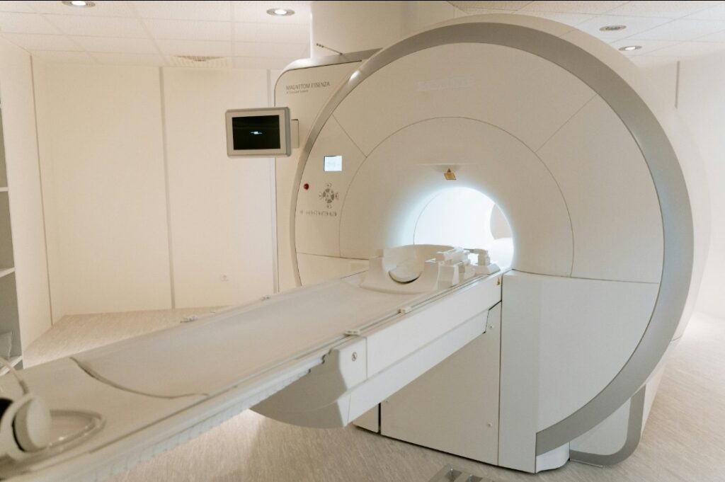
What is a CT scan?
The full name of CT scan is computed tomography. CT scan is a multiple rotating pencil beam of X-rays processed by a computer and a detector arranged in a complete circle around patient. The reconstruction of these areas on 2 – D, dimensional display provides characteristic CT scan appearance.
Computed axial tomography(CAT) is a synonym for CT. It refers to the axial plan, the most common plan for CT scans.
The spiral or helical CT scanners use a large bank of detectors ( multislice ) and the patient moves through the field during scanning so that the X-ray beams describe a helical path.
How does a CT scan work?
A CT scan uses an X-ray to produce an image. The same basic principle for CT scans as for all other X-ray studies. X-rays are blocked by tissues depending on their density ( atomic number ). Air is black on CT and mineral is white.
To obtain the slice, an X-ray source is around the body in an arc while an X-ray detection source rotates opposite the source around the opposite side of the body.
History of CT scan
The first ever CT scan was taken on October 1, 1971, by Dr. James Ambrose at Atkinson Morley Hospital on a middle-aged female patient who was suspected of a tumor in the left frontal lobe. A CT scan found as a very important and useful tool in medical treatment. The contributions of Godfrey Hounsfield and Allan Cormack. However, other researchers worked in areas related to CT for the discovery and development in the next decade or two . Godfrey Hounsfield developed his first CT system after years of struggles he simplified the problems of two dimensional into series of parallel slices . On November 1967 to July 1968, and it was during this period the idea for a “3D x-ray” was formed.2 By June.
Which Part of the body is most studied with a CT scan?
A CT scan can be used from head to toe but it is majorly used for:
- Head; such as cranial trauma, Suspected internal bleeding, or stroke.
- Neck
- Facial bones
- Chest
- Abdomen
- Pelvis
- Knee; Evalute Tibial Plateau fracture and ligament injury etc.
- Hip; Specifically for assessment of acetabulum fracture.
- Calcaneus; Fracture
What is a contrast CT scan?
Contrast CT scan is a method used for vascular structure. Dense substances such as bone can be seen easily in X-rays but the soft tissues are not visual as they stop the X-rays so we use an agent. In this iodine (Mineral density ) is injected intravenously. It helps to identify the vascular structure and can display the difference between normal from abnormal structures. Contrast CT scans also help to identify certain types of pathology such as infection(abscess), malformation and metastatic cancer, or aneurysm. The barium component is also used as a contrast agent for imaging the digestive system.
How to prepare for a CT scan?
Before doing a CT scan some instructions are made, such as:
Clothing : The person should change into patient gown. Remove all the jewelry and valuable products at home or before entering the scanning rooms.
Allergy: Please share the details of the allergic substance with your consultation doctor. If the patient has an allergy to contrast media then a CT scan is prohibited. Be cautious about discussing these details before going to the scanning room. If you have any reaction to contrast media then immediately inform the Physician.
Eating and Drinking: If you are having a CT scan then you can eat and drink normally but if you are taking a contrast CT scan then eating and drinking is restricted before 3 hours.
Medication: The patient can take the prescribed medication as usual.
Pregnancy: After consultation from doctor you can do Exam .
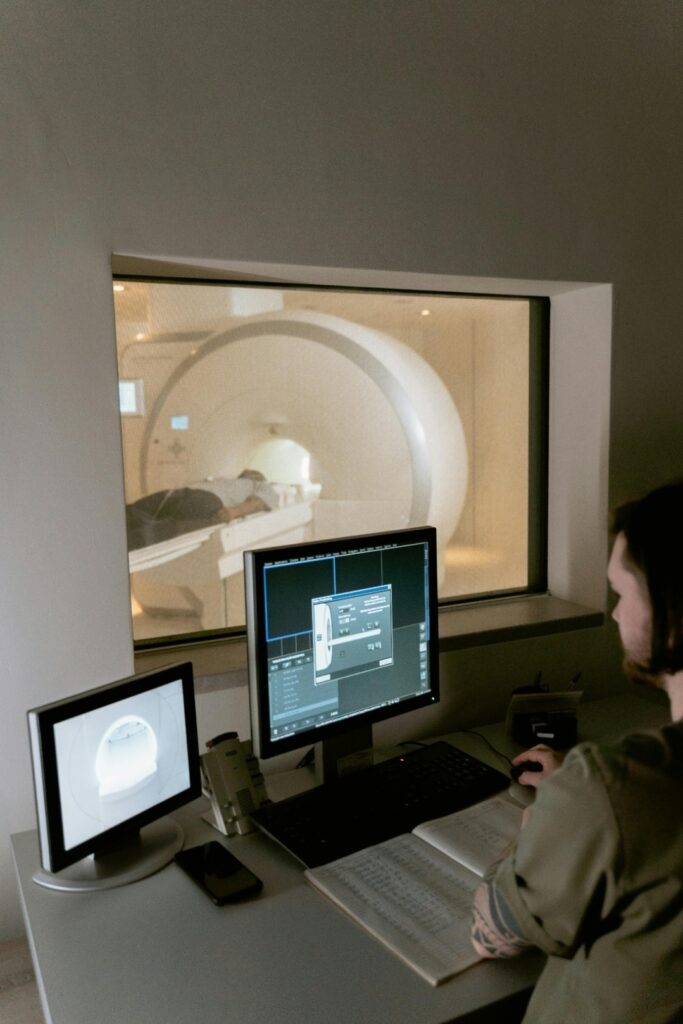
What Happens in CT scan?
During CT scan
- Before entering to scanning room the person should change into the patient gown and remove all the valuables. You can places your personal belongings in a locker.
- In contrast CT scan, the person is given contrast media intravenously or orally as a liquid.
- You will lie on the scan table still with no movement.
- The technologist will be in another room. He /she will communicate with the speaker present in the scanning room. There will be a call button for your safety reasons if you have any problem regarding anything then you can press it to call technologist for help.
- The machine starts to rotate around your body and an X-ray beam will enter your body for a short while. During this process, you will hear a normal clicking sound.
- You can feel suffocation, sweating, and numbness. If you have taken contrast media then you can feel a flushing sensation, metallic taste in the mouth, headache, nausea, and/or vomiting.
- After completion of the IV line which was inserted for contrast media, the line will be removed.
After CT scan
- Side effect of contrast media is a common problem faced by patients. A short period of observation is needed, notify us if you have any side effects or reactions.
- You can get back to your normal activities as soon as the exam is over.
- Sometimes doctors can give specific instructions after the procedure, depending on the particular situation.
Advance in CT technology
Spinal CT scan
If an MRI is unavailable, a CT of the spine can demonstrate the bony canal, intervertebral foramen, and disc protrusion. After instilling some intrathecal contrast, CT scanning clearly demonstrated lesions compressing the spinal cord or the cervical-medullary junction.
Coronal CT scan
In the absence of the latest scanners and good reconstruction, full neck extension combined with maximal angulation of the CT gantry permits direct coronal scanning. A coronal CT -scan shows a tumor of the ethmoidal sinus.
CT angiography
Helical scanning during infusion of intravenous contrast provides a non-invasive method of demonstrating intracranial vessels in 2 and 3-D CT angiography is as accurate as conventional angiography in detecting small aneurysms.
CT scan in bone diseases
Computed tomography (CT) is used selectively for assessing patients with bone and joint disease. CT may be used when skeletal configuration needs defining when calcific lesions are being assessed, when MRI is contraindicated, or when articular regions are being evaluated in which an adjacent joint replacement creates signal artifacts on MRI, using specific metal artifact reduction algorithms.
CT scan for brain
CT – scan is good for demonstrating bone and calcification well. It will also detect abnormalities of the brain and ventricles, such as atrophy, tumors, cysts, abscesses, vascular lesions, and hydrocephalus but a CT – scan is not optimal for lesions of meninges, and cranial nerves.
CT scan in Respiratory condition
Computed tomography (CT scan ) provides detailed images of the pulmonary parenchyma, mediastinum, pleura, and bony structures. The displayed range of densities can be adjusted to highlight different structures, such as the lung parenchyma, the mediastinal vascular structures, or the bone.
In cases of suspected lung cancer, CT is central to both diagnosis and staging and facilitates percutaneous needle biopsy.
CT pulmonary angiography (CTPA) has become the investigation of choice in the diagnosis of pulmonary.
CT may assist in identifying the cavitation of tuberculosis, fungal infection
, and other signs of infection (halo–air crescent) thromboembolism. CT may be used to assess disease progression, thereby predicting prognosis and in screening to detect the earliest signs of disease.
Computed tomography angiography (CTA)
Computed tomography angiography is a computed reconstruction of vascular structures in the body. In CTA, iodine is injected using a power injector for both venous and arterial imaging. CTA is used to detect thrombosis within veins or arteries, stenosis, aneurysms, vascular malformation, and pulmonary embolism.
Computed tomography urography (CTU)
Computed tomography urography (CTU) is used to evaluate cysts and mass lesions in the kidney or filling defects within the collecting systems. CTU has largely replaced the previous gold-standard investigation of intravenous urography (IVU) for the investigation of the upper urinary tract.
For the investigation of patients with renal trauma, a triple-phase CT scan with a delayed phase, to assess the integrity of the collecting system, is performed.
What are the benefits of a CT scan compared to an MRI?
A CT scan is generally quicker, less expensive, and readily available than MRI. It is often better than MRI at assessing cortical bone and for specific density measurement. Please note there is variation in the type and quality of imaging depending on department, training, the expertise of the radiologists, and the preference of clinical staff.
Recent Post
How to Cure Insomnia Naturally ?
How to Cure Insomnia Naturally If you’re struggling with insomnia, the good news is that effective treatment doesn’t always require medication. Research shows that several

Understanding your back problems at once
Understanding your back Problems at once Introduction Because you clicked on this article, it is likely that you have had firsthand experience of how frustrating
What is the best diet to lower LDL cholesterol?
What is the best diet to lower LDL cholesterol? Introduction If you are concerned about high cholesterol, especially elevated LDL (Low-Density Lipoprotein) cholesterol, making the
What is best diet for high cholesterol ?
What is best diet for high cholesterol ? Maintaining healthy cholesterol levels is essential for protecting the heart and reducing the risk of cardiovascular

Best 25 Heart Healthy Foods to Eat
Best 25 Heart Healthy Foods to Eat Heart disease remains one of the leading health concerns worldwide, but many risk factors can be managed through
Sharp Pain in the Heel of My Foot: Causes and Treatment
Sharp Pain in the Heel of My Foot: Causes and Treatment Pain in the heel of my foot is one of the most common foot